
Table of Contents
Name of Procedure
- Knee joint intra-articular steroid injection
- Supartz/synvisc/orthovisc/euflexxa/hyaluronic acid knee injection
- Viscosupplementation
Sample Opnote
Goal
To inject a medication into the knee joint.
Indications
Depending on what’s injected, usually it is to treat pain in the knee from a variety of etiologies such as osteoarthritis.
Contraindications
- Common contraindications
- Potentially hardware in or around the joint, depending on the procedure.
Anatomy
For the purposes of a knee injection, knee anatomy is straightforward. You simply need to find the space between the femur and tibia. Other soft tissue anatomy generally doesn’t effect the procedure.

Equipment/Skills/Setup
- C-Arm
- Bed for C-Arm
- 25g x 1.5″ hypodermic needle
- Contrast
- 1 or 2 3cc syringes (may not need as noted below) filled with desired injectate:
- bupivacaine/lidocaine
- supartz/synvisc/orthovisc
- steroid (dexamethasone/triamcinolone)
- Prep solution
- Drape to keep injection site clean
Landmarks and Patient Positioning
- Position the patient in a basic supine position so that the bottom of the c-arm can go under the table below the knee.
- Bend the knees roughly 90 degrees and put a roll under it so the patient’s legs can stay relaxed. This opens up the joint space a bit. Having someone help hold the patient’s foot can be helpful.
Technique
- Position the C-arm in an AP orientation with the top angled slightly towards the feet.
- Playing with the angle will allow you to see which opens up the joint space the best.
- Use a pointer to find a spot directly over the joint space. We often choose just lateral or medial to the patellar ligament.
- Insert a 25g x 1.5″ hypodermic needle in a straight AP direction towards the joint space (“straight down the barrel”).
- As seen in the image, even if not directly over the joint space you can simply aim for an opening to enter the space, often in the middle of the knee.
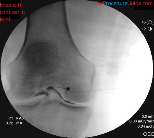
- Inject a small amount of contrast. Ideally you see spread along the joint as in the image. A simple “blob” of contrast may mean that the needle tip is in the subcutaneous space.
- Inject the desired medication based on the specific procedure that you’re doing.
Tips
- Usually a 25g x 1.5″ hypodermic needle is sufficient to enter the joint space. With this approach no second/larger needle is needed,.
- As a result, it can be done without local anesthetic since it’s the same needle that would be used to administer local anesthetic anyway.
- If it turns out that the hypodermic needle is not long enough, just inject lidocaine through it while pulling out.
- Then use a longer needle such as a bent quincke needle to reach the joint space.